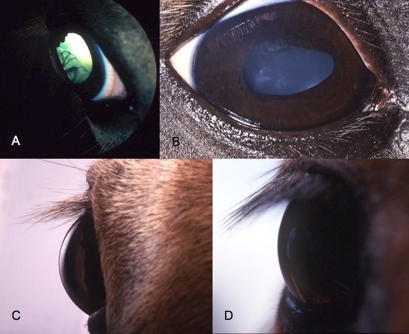
Clinical signs of MCOA syndrome.*
In this condition, certain parts of the eye, such as the cornea, iris, lens, ciliary body, and retina, do not develop normally. One of the most common and apparent abnormalities is an enlarged cornea that gives the eyes a “bugged-out” look. Horses with ASD are at increased risk for vision loss.
If you are intending to breed a horse, the ASD gene status of your horse is something you should know in order to select a mate that will minimize chances of a homozygous ASD foal, as it is only the homozygous foal that has any chance of having affected vision.
Generally,
this condition does not cause blindness but severely afflicted horses might have
cataracts and some reduction in sight.
In the Miniature Horse breeds, the color known as “chocolate” with a flaxen mane
and tail is very popular, and has become strongly associated with this horse.
The “chocolate” color is also found in other breeds, most commonly in Icelandics,
Shetland Ponies, Miniature Horses and some draft breeds.
The chocolate color is caused by a color modifying gene – that is, a gene which
modifies (in this case dilutes) another color. This gene has been named the
“Silver Dapple gene” by geneticists. It is a gene that only dilutes the color
black. It does not affect the color red (sorrel or chestnut). Therefore, a
sorrel or chestnut horse can carry the gene and pass it on, but you cannot tell
whether the horse carries the gene from looking at the horse, as there is no
black on its body to modify.
The silver dapple gene is a dominant gene which produces an even more dilute
effect if two of them are present in the animal (one inherited from each
parent). In these homozygous horses, the manes and tails are almost white, and
the black on the body is lightened up to an almost gray color. If it is a “red
chocolate” horse (silver dapple bay), the black “points” on the legs are so
light that horse almost looks like a solid chestnut with a white mane and tail,
at least when the horse is young.
The abnormal ASD gene is a “semi-dominant” gene. When the horse inherits only one abnormal gene from one of its parents, there is only the very slight and harmless physical indication of cysts (or none if it is a silent carrier) behind the lens. Cysts can usually only be detected by an experienced veterinary ophthalmologist using an indirect ophthalmoscope. About 87% of horses that carry a single ASD gene will show “cysts.” The remaining 13% are “silent carriers,” and can pass on the gene to offspring to the same degree as horses whose genetic status is visible in an eye exam - that is, 50% of the time.
When a horse inherits two of the dominant form ASD genes, one from each parent, the eyes of the horse will almost always show a variety of other differences in addition to the cysts. (This is the horse that in lay terms has become known as the “ASD” horse.) Many of these are easily seen if you know what to look for. Not all homozygous horses will have all of the various differences which are part of the ASD syndrome – most will exhibit only some of them. Common differences which can be seen without special equipment include megalocornia (a more steeply curved cornea than normal - "pop eyes"), and a pupil with an abnormal shape. One difference that is almost diagnostic is the inability of the pupils of the homozygous horse to dilate or dilate normally, following administration of drugs used for this purpose.
Again....If you are intending to breed a horse, the ASD gene status of your horse is something you should know in order to select a mate that will minimize chances of a homozygous ASD foal, as it is only the homozygous foal that has any chance of having affected vision.
The horse
Silver dilution gene dilutes black pigment but has no effect on red pigment. The
mane and tail are lightened to flaxen or silver gray, and may darken on some
horses as they age. A solid black horse with this gene will be chocolate colored
with a lightened mane and tail. A bay horse will have the black pigment on the
lower legs, mane and tail lightened. Sometimes bay horses with Silver dilution
can be mistaken for chestnuts with a flaxen mane and tail. Silver dilution is
inherited as a dominant trait. It is known to occur in Rocky Mountain horses and
related breeds, Shetland ponies, Icelandic and Morgan horses.
The gene responsible for Silver dilution has been identified as PMEL17 with a
mutation in exon 11 being responsible for the dilute phenotype described above.
Research has also confirmed the Silver dilution mutation to be associated with
Multiple Congenital Ocular Abnormalities syndrome (MCOA), a wide range of ocular
defects occurring in the anterior and posterior segment of the eye. The severity
of the syndrome is dosage related, thus horses with 1 copy of Silver have less
severe signs than those with 2 copies of the mutation.
To avoid producing
offspring with severe MCOA, breeders should not breed 2 Silver dilute horses
together.
Silver Dilution results are reported as:
N/N No copies of Silver dilution detected.
N/Z One copy of Silver dilution detected.
Z/Z Two copies of Silver dilution detected. Horse is expected to have MCOA
abnormalities.
You can read more about this and submit your horses dna here for testing.
Do you
have a topic you would like to see here or article you'd like to submit?
Please
E-mail me
We wish to thank all of the people that have shared their experiences &
knowledge with others by sending us tips and donating articles. For articles on
our pages that have been reproduced, credit is given and a link is posted to
your site (reproducing articles archives information on our server which
decreases broken links that occur when content on the site was either removed or
relocated without proper redirects in place to lead visitors to the correct
page.)
Please forgive us if we did not give full credit for any one of the wonderful
articles here. If we have used something of yours that you would like removed
please let me know.
All rights reserved. No part of any pages may be reproduced in any form or
means without written permission of Lil Beginnings Miniature Horses.
Lil Beginnings Miniature Horses is not responsible for any death, injuries,
loss, or other damages which may result from the use of the information in our
pages.
ASD - Silver Gene in Miniature horses and Shetland ponies - Silver dilution mutation